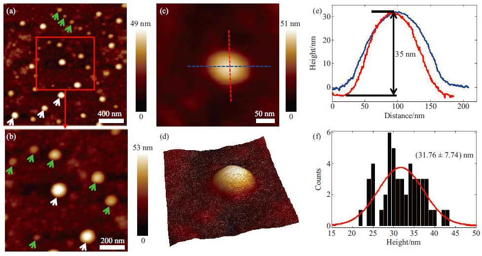
AFM in situ imaging visualizing single living exosomes immobilized on substrates. (Image by SIACAS)
Recently, researchers from Shenyang Institute of Automation Chinese Academy of Sciences (SIACAS) have successfully utilized atomic force microscopy (AFM) to probe the native behaviors of single living exosomes under aqueous conditions. Related study is published as a cover paper in Progress in Biochemistry and Biophysics with the title Nanostructures and mechanics of living exosomes probed by atomic force microscopy.
Exosomes are cell-secreted extracellular vesicles with a size range of 40 to 160 nm in diameter and exosomes contain various biomolecules to regulate the physiological/pathological processes. Investigating the structures and behaviors of individual exosomes is of critical significance for understanding the biology of exosomes and their practical applications. Nevertheless, due to the lack of adequate tools and methods, the detailed structures and mechanics of living exosomes in their native states are still not fully understood.
In this work, the researchers presented an immobilization method based on electrostatic adsorption for attaching living exosomes onto the substrate, which allowed obtaining the topography of single living exosomes with high quality under aqueous conditions by AFM. Besides, the mechanical properties of single living exosomes were quantified and visualized by AFM.
The study provides a novel way for probing the native behaviors of living exosomes at the nanoscale, which is of general significance for life sciences.
CONTACT
LI Mi
Email: limi@sia.cn