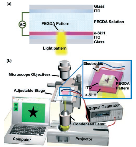
Illustration of the experimental system and the OEK device.(a) The two-dimensional structure of the OEK chip. (b) The experimental system consists of a computer, a signal generator, a display projector, and a microscope; the inset figure details the different layer structures of the OEK device. (Image provided by LIU Na et.al)
The culturing of cancer cells on micropatterned substrates can provide insight into the factors of the extracellular environment that enable the control of cell growth. Researchers from Shenyang Institute of Automation (SIA), the Chinese Academy of Sciences (CAS), Department of Mechanical and Biomedical Engineering, City University of Hong Kong, and Department of Power Mechanical Engineering, National Tsing Hua University report here a novel non-UV-based technique to quickly micropattern a poly-(ethylene) glycol diacrylate (PEGDA)-based hydrogel on top of modified glass substrates, which were then used to control the growth patterns of breast cancer cells. Previously, the fabrication of micropatterned substrates required relatively complicated steps, which made it impractical for researchers to rapidly and systematically investigate the effects of different cell growth patterns.
The technique presented herein operates on the principle of optically-induced electrokinetics (OEKs) and uses computer-generated projection light patterns to dynamically pattern the hydrogel on a hydrogenated amorphous silicon (a-Si: H) thin-film, atop an indium tin oxide (ITO) glass substrate. This technique allows us to pattern lines, circles, pentagons, and more complex shapes in the hydrogel with line widths below 3 mu m and thicknesses of up to 6 mu m within 8 s by simply controlling the projected illumination pattern and applying an appropriate AC voltage between the two ITO glass substrates. After separating the glass substrates to expose the patterned hydrogel, we experimentally demonstrate that MCF-7 breast cancer cells will adhere to the bare a-Si:H surface, but not to the hydrogel patterned in various geometric shapes and sizes. Theoretical analysis and finite-element model simulations reveal that the dominant OEK forces in our technique are the dielectrophoresis (DEP) force and the electro-osmosis force, which enhance the photo-initiated cross-linking reaction in the hydrogel. Our preliminary cultures of breast cancer cells demonstrate that this reported technique could be applied to effectively confine the growth of cancer cells on a-Si:H surfaces and affect individual cell geometry during their growth.
This work was published on Lab on a Chip (2014) 14, pp: 1367—1376.
Contact:
LIU Lianqing
Email: lqliu@sia.cn
Shenyang Institute of Automation