High-resolution imaging of living cell at the micro-/nano-scale is important for life science research. It may help to observe biological activities of cells, and to detect cell responses to external stimuli and even movements of some protein molecules in cell membranes. However, effective methods to realize such goals do not exist.
Scanning ion conductance microscopy (SICM), first introduced by Hansma group and further developed by korchev group, has become a promising tool for high-resolution imaging on biological samples. By measuring the ion conductance change through an electrolyte-filled fine tapered glass nanopipette, the tip-sample distance can be controlled and maintained thus to obtain the topographical images of samples. Unlike other scanning probe microscopy (SPM) techniques that require tip-sample contact, SICM is naturally a non-contact approach. Therefore, it can be used to probe live cells under physiological conditions, which is difficult by other SPMs that can easily modify and damage the soft living cells. There are at least two imaging modes of SICM: continuous mode and hopping mode. In both imaging modes, the low imaging speed is the common disadvantage that hinders the efficiency of SICM based observation.
To solve this problem, researchers from Shenyang Institute of Automation (SIA), Chinese Academy of Sciences developed a prior knowledge based fast imaging method (as shown in Fig. 1) based on home-built scanning ion conductance microscopy recently. The key idea is to achieve a high resolution image on targeted features by scanning the sample twice, the first scan to obtain prior knowledge on targeted features with low resolution and the second scan to obtain images on targeted features with high resolution. In the first scan, the scanning speed can be very fast because the prior knowledge can be obtained from a low resolution image that requires few scanning points. In the second scan, only the interested areas (targeted features) are scanned. Because the scanning area on targeted features is much smaller than the whole scanning region, it takes much less time to obtain high resolution images on targeted features, As a result, the total time to acquire a high resolution is much less than the conventional one scan method .
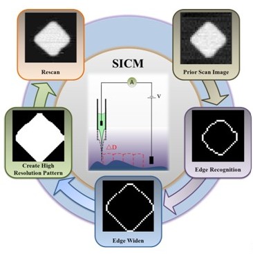
Fig. 1 Flowchart of fast imaging method.(Image provided by LI Peng et,al.)
To verify the effectiveness of this method, the researchers firstly constructed a home-made SICM according to the setups shown in Fig. 2. Sample is moving up and down driven by the nano stage that is extending and retracting. The ion current, varying with respect to the distan- ce between the nanopipette tip and the sample surface, provides an input signal for feedback control system to adjust the vertical axis of nano stage and ensures that the sample and nanopipette tip are out of contact.
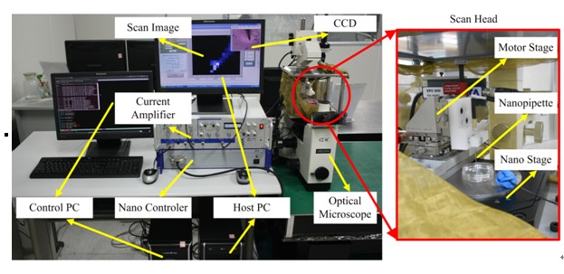
Fig. 2 Photograph of SICM (Image provided by LI Peng et.al,)
Then, the researchers applied the proposed fast imaging method to image polydimethylsiloxane (PDMS) samples in Fig. 3. Compared with the low resolution prior images, these reconstructed images based on the fast imaging method show similar but more subtle topography. According to TABLE I. scan pixel counts of both low resolution and high resolution images of the PDMS grating are 78% of full scan counts. And The scan time is consuming 70% of full scan time, Though there is not significant time saving in the 64 ´ 64 pixels fast imaging, using the same prior scan image of 32´ 32 pixels, higher resolution image (more than 64´ 64) pixels would be more advantage in time saving. And it is also more advantages when the features are smaller.
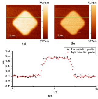
Fig. 3 Experimental results of PDMS grating obtained by SICM with normal scanning and Fast imaging method. (a):32 ´ 32 pixels of SICM low resolution image got by normal full scan; (b) : 64 ´ 64 pixels of high resolution reconstructed images based on Fast imaging method is guided by (a).(c):line profiles analysis are based on the lines indicated in the upper images. (Image provided by LI Peng et.al)
TABLE I. Comparison of the scanned results
|
|
PDMS Grating |
|
|
Scan points counts |
Scan time (s) |
|
Traditional full scan |
4096 |
235(´4) |
|
Fast imaging (for both images) |
3175 |
656 |
|
Ratio(%) |
78% |
70% |
This work was published on 2013 IEEE/ASME International Conference on Advanced Intelligent Mechatronics (AIM), pages 89 – 93, 9-12 July 2013. It was supported by the National Natural Science Foundation of China (No. 61175103), the Instrument Developing Project of the Chinese Academy of Sciences, Grant No. YZ201245 and the CAS/SAFEA International Partnership Program for Creative Research Teams.
CONTACT:
LI Peng
Shenyang Institute of Automation
Chinese Academy of Sciences